Hidez Science
Muscle Structure
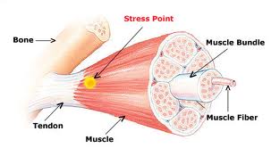
Whether in horses or in humans,each muscle consists of thousands of cells that are bundled together to form one functional unit. Skeletal muscles are covered with a protective sheath that eventually comes together to form tendons & ligaments. Muscles have a plentiful blood supply, because they require constant delivery of oxygen and nutrients. The blood supply should also take away the toxic waste substances that muscles build up during their high levels of activity.
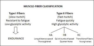
Equine, muscle fibers are classified as either slow twitch or fast twitch fibers. Slow twitch or Type I fibers (which are white in the stained muscle section pictured) are highly oxidative, meaning they use aerobic metabolism to produce energy-generating ATP. These fibers are use for endurance they are "fatigue-resistant" as they are capable of reducing the toxic end products such as lactate or lactic acids.
Fast twitch fibers or Type II, fibers are subdivided into Type II A (stained a dark tan colour) & Type II B (stained a lighter tan color) fibers.
Type II A fibers are both high & low oxidative. These fibers are capable of utilizing both aerobic metabolism to produce energy for work & are used to maintain high speed or for jumping.
Type II B fibers are low oxidative, meaning they are highly anaerobic & are used to give horses speed. Neither class of Type II muscle fibers has the ability to reduce lactate as do Type I fibers, meaning fatigue is reached in a shorter time.
Distinct differences exist in the ratio of Type I to Type II fibers among breeds of horses, more specifically among type of preformance. Quarter horses & Thoroughbreds have lower proportion of Type I fibers when compared to Arabians or Andalusians.
Muscle Energy Sources
All cells are reliant upon high-energy phosphate bonds for energy. When these powerful phosphate chemical bonds breakdown, they release energy which is used by the body to power thing, like pumps, enzymes, and revolving doors for proteins to come & go from cells.
The fastest method of producing energy comes from a molecule that stores phosphate, know as creatine phoshate or phosphocreatine. However, this hot fuel called creatine phospate or phosphocreatine only lasts for less than 1 minute.
For more continuos function muscle will be need to make & store a more reliable energy source. The most 'reliable' energy is found in a molecule called ATP. It's so important it's often called "energy currency." ATP itself must come from the breakdown of nutrients-sugar (glucose, or Glycogen in it's stored form), fats & protein. ATP is continually used, remade & reused again. If there is no ATP the muscle will not fuction.
How Muscle Act
Muscles act by using ATP (adenosine tri-phosphate) to bind to different muscle fibres to cause a contraction of the whole fibre bundle. This process releases a phosphate ion from ATP to made ADP (adenosine di-phosphate). ADP has the phosphate ion put back on by the breakdown of sugar in the muscle cell and from stores in the muscle as creatine phosphate. The release of the phosphate ion from creatine phosphate is controlled by the enzyme creatine kinase (CK).
ADP and ATP are in relatively small concentration in muscle cells. Conversely, CK is in very high concentrations and is rapidly depleted by intense short term exercise, such as sprints, whilst ATP/ATP concentrations remain stable.
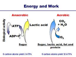
ATP is basically raw energy to all cells. ATP is the end product of two connected processes: anaerobic and aerobic metabolism. Anaerobic metabolism is the rapid breakdown of sugar to essentially lactic acid. Aerobic metabolism, a slower process, is the breakdown of lactic acid to carbon dioxide, which, unlike lactic acid, can be breathed out of the system.
Therefore, if you work hard, you will max out your aerobic system and start producing lactic acid. The build-up of acid in the muscle will eventually cause cramps and stop you working.
When muscles work hard and are overstretched, fibres break and the membranes leak. One of the enzymes that leak into the blood in high concentrations is CK. Therefore a good marker of muscle injury is CK.
REFERENCES
1. MURRAY STEWART,Muscle Structure and Function- an explanation, Equine Vet. Journal (1976)
2. SEEHERMAN,M.W. O'CALLAGHAN, S.H. SCHELLING, MARY ROSE PARADID and R.S. STECKEL, Scintigraphic identification of skeletal muscle damage inhorse 24 hours afterstrenuous exercise, Equine Vet. Journal (1991)
3. JENNIFER M. MACLEAY, JESSICA A. BILLSTROM, MELISA A. HOWER-MORITZ and MICKELSON, Skeletal muscle metabolic response to exercise in horses with 'tying-up' due topolysaccharide storage myopathy Equine Vet. Journal (1999)
4. K.Leisson, U., Jaakma, T. Seene, Adaptation of locomotor muscle fiber types to endurance and intensive high speed training, Journal of equine Vet. Science (2008).
5. B. Eessen-Gustavsson and S. Valberg, Blood and Muscle Ammonia Concentrations in Horses during Treadmill Work and after Racing. Equine Exersice Physiology, 1987.
Cutmore,C.M.M.,Snow,D.H. and Newsholme,E.A. (1986). Effects of training on enzyme activities involved in purine nucleotide metabolism in Thoroughbred horses. Equine Vet.
6. Snow, D.H., Harris, R.C.and Gash, S.P. (1985. Metabolic response of equine skeletal muscle to intermittent maximal exercise. J.Appl. Physiol
Saltin,B. and Gollnick,P.D. (1983) Skeletal muscle adaptability: Significance for metabolism and performance, handbook of Physiology
7. NAT Brown, CE Kawcak, Architectural properties of distal forelimb muscles in horses, Equss caballus, Journal of Morphology.